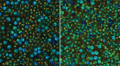
Biomedical imaging at Morgridge comprises a dedicated group of technology innovators who develop imaging, computation, and fabrication tools for understanding and solving biomedical problems. Our investigators are broadly interested in multiscale, label-free, quantum, and quantitative and other approaches that shed light on currently unseeable or poorly understood biology. As a hub for imaging on campus, we collaborate with leaders across biomedical sciences at Morgridge, UW–Madison, and beyond whose questions help us further push the technology to new capabilities.
Investigators
Melissa Skala’s lab develops new methods to understand and combat cancer using photonics-based technologies. Optical technologies are attractive for clinical translation because these techniques are inexpensive, portable, and fast — providing a wealth of information on tissue structure and function. We are particularly interested in developing personalized cancer treatment strategies, and in developing more effective cancer therapies. Technology development focuses on metabolic and functional imaging of the tumor and its microenvironment (e.g. metabolic activity, blood vessel morphology, blood oxygenation, blood flow, and molecular expression).
The Randy Bartels Lab develops new imaging technologies to expand the frontiers of biomedical research and explore new biological questions. Bartels and his team employ a focus on optical imaging due to its unique ability to resolve spatial scales ranging from sub-cellular structures such as organelles to large regions of tissue. Critically, microscopic structure elucidation allows us to understand morphological changes associated with disease. Due to a diverse range of biological unknowns, current projects utilize a unique swath of techniques — from optical and quantum physics approaches to microscopy, advanced computations and algorithms, and translational biology.
Juan Caceido’s lab utilizes the power of machine learning to illuminate the biological secrets hidden within microscopy images of cells and tissues. These microscopic glimpses can be used to infer the biological properties of the underlying organism. The development of novel computational techniques make the phenotypic information of these images readily available, without the influences of noise and unwanted variation. Training of the next CHAMMI-75 model — a large and highly diverse curated dataset of multi-channel microscopy images designed to advance foundation models for cellular biology — is currently under way, further quantifying cellular morphology and making it useful for biological studies.
Kevin Eliceiri is the director of UW–Madison’s Center for Quantitative Cell Imaging (CQCI) — an umbrella entity for the Eliceiri group and its sister biological optics lab run by Cell and Regenerative Biology Assistant Professor Abhishek Kumar. Research at CQCI investigates light interactions with biology, or biophotonics. The CQCI is located in the Laboratory for Optical and Computational Instrumentation (LOCI) in the Animal Sciences Building on campus. Here, instrument innovators at Morgridge have a second home.
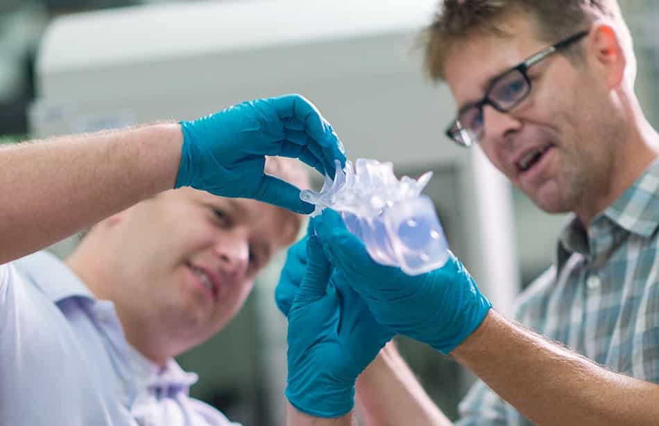
The Fab Lab — a collaborative space led by Eliceiri that specializes in device design, 3D fabrication, and microfluidic production — is available to UW and Morgridge investigators who wish to work on new designs for biomedical research and clinical use. Current key projects include a novel imaging and wounding device for studying wound healing in zebrafish, a new multiscale imaging instrument that combines optical and ultrasound approaches to characterize breast cancer density and an open source tomographic light sheet microscope for understanding morphological changes in developing tissue.
Eliceiri is also the co-chair of Bioimaging North America (BINA). This organization (housed at Morgridge) connects imaging scientists from across the continent to accelerate their growth and foster collaboration. In 2024, Madison hosted the BINA Community Congress international imaging meeting, with over 300 attendees, and will return as host in 2026.